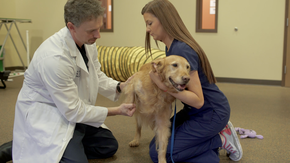
Performing a systematic physical examination and visual observation of the patient’s mobility will provide an initial assessment on where the potential issue(s) may exist. A thorough orthopedic exam allows for palpation of the limbs and joints while moving them through their range of motion to draw attention to changes within the tissue and help assess if the patient seems painful. Additionally, it is important to complete a neurologic evaluation and consider running additional diagnostic tests to look for any underlying disease processes that could affect treatment options.
Pain Evaluation
According to the Summary of 2015 AAHA/AAFP Pain Management Guidelines for Dogs and Cats, the most accurate method for evaluating pain is through observing any changes in the pet’s behavior. A pain score is considered the fourth vital sign after temperature, pulse and respiration.1 A patient’s pain should be evaluated in the clinic and by the pet owner to paint a full picture of how the pet is acting.
Pet Owner
Feedback from the owner is vital in understanding where, the length of time and severity of how the pet has been affected to determine the next steps for evaluating if arthritis is present. He or she should be asked a series of questions about any changes in behavior and given a pain scale form to fill out based on whether it is an acute or chronic problem. This will provide valuable insight into the pet’s everyday activities and how they vary.
Standing Evaluation

Gathering data on how a patient is bearing weight while standing can provide objective numbers to determine which limb or limbs are effected. It has been shown that bathroom scales can be used as a reliable way to measure static weight bearing in canines2; therefore, a free-standing platform with built-in sensors, such as the Stance Analyzer, can visually show where a potential lameness exists. This sort of tool takes up minimal floor space and can be a cost-effective option in helping obtain a diagnosis, as well as measuring patient outcomes. Recording this sort of information can help guide treatment plans and provide owners with a better understanding of the source of their pets’ complications.
Gait Analysis
Objectively measuring a patient’s gait can be performed via force plate or a commercially available portable walkway. Force plate is considered the gold standard for evaluating gait by calculating the ground reactive forces of a cat or dog while standing, walking or jogging over the plate. The portable walkway is a mat that allows a pet to stand, walk or jog providing information on gait symmetry, stride length and the amount of pressure placed on each paw. These types of devices are expensive and take up a lot of floor space, thus they are not as common of an option for general practice.
Imaging
It is very important to obtain a picture of the joints suspected of having osteoarthritis. Radiographs are most commonly taken to assess the bony changes associated with OA. MRI and CT offer additional diagnostic capabilities, though less commonly used to assess OA, with MRI showing how the soft tissues are involved, while CT can offer better detail for the bony changes in more complex joints.
Arthrocentesis
Collecting a sample of the fluid in a swollen joint or joints suspected of having arthritis can help rule out what is causing the swelling or pain, especially in younger patients, or in those with a more complicated clinical history. The color, consistency and cellular make-up of the synovial fluid can provide clues as to what changes are occurring within the joint and why.3
References
1. Epstein, M. et. al. (2015). 2015 AAHA/AAFP Pain Management Guidelines for Dogs and Cats. JAAHA, 51(2), 67-84. doi: http://dx.doi.org/10.5326/JAAHA-MS-7331.
2. Hyytiäinen, H.K. et. al. (2012). Use of bathroom scales in measuring asymmetry of hindlimb static weight bearing in dogs with osteoarthritis. Veterinary and Comparative Orthopaedics and Traumatology, 25(5), 390-396. doi: https://doi.org/10.3415/VCOT-11-09-0135.
3. Degner, D. (2014, August). Arthrocentesis in Dogs. Clinician’s Brief. Retrieved from https://www.cliniciansbrief.com/article/arthrocentesis-dogs.
